[vc_row][vc_column][vc_column_text]Thank you to KITV for running a story on the evening news of our mission here in Hawaii.
[/vc_column_text][/vc_column][/vc_row][vc_row][vc_column][gem_button position=”center” size=”medium” corner=”3″ icon_pack=”elegant” text=”See video on KITV” link=”url:https%3A%2F%2Fwww.kitv.com%2Fstory%2F39608429%2Fcoral-showing-abnormal-growth-in-kaneohe-bay-could-be-cancerous||target:%20_blank|”][/vc_column][/vc_row]
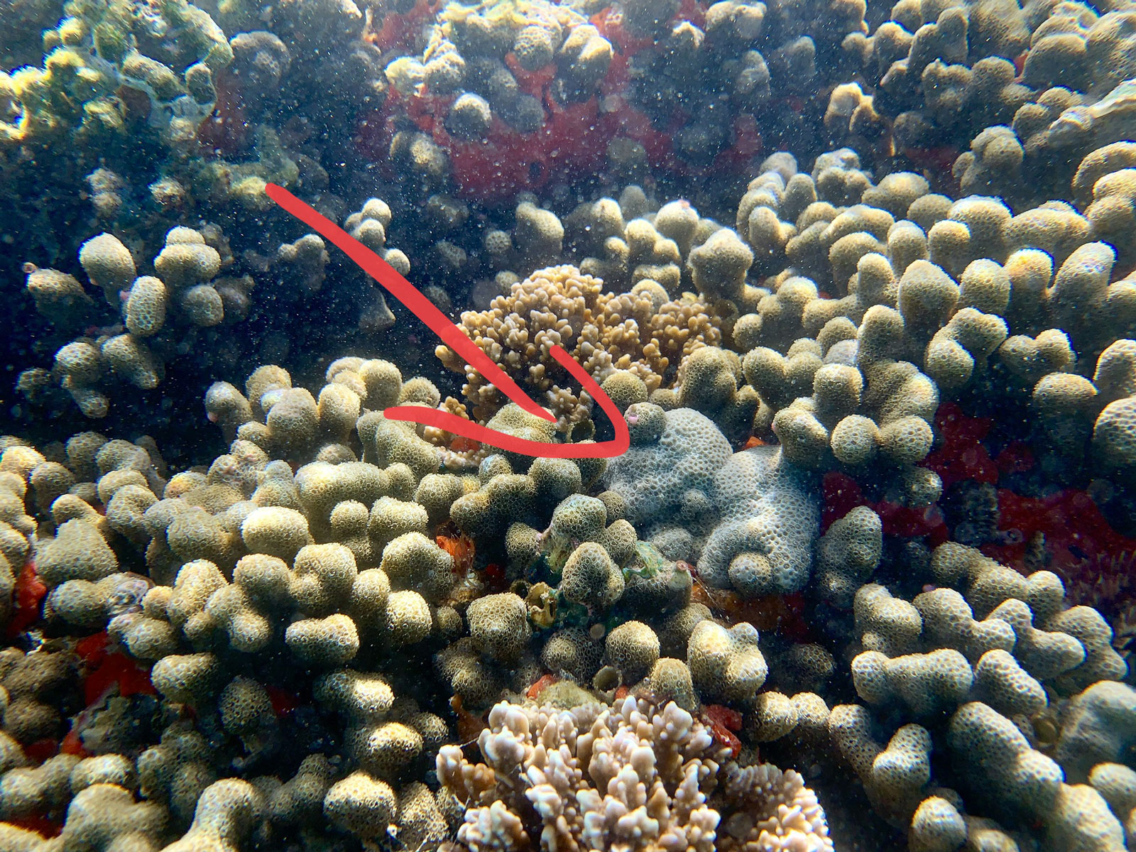
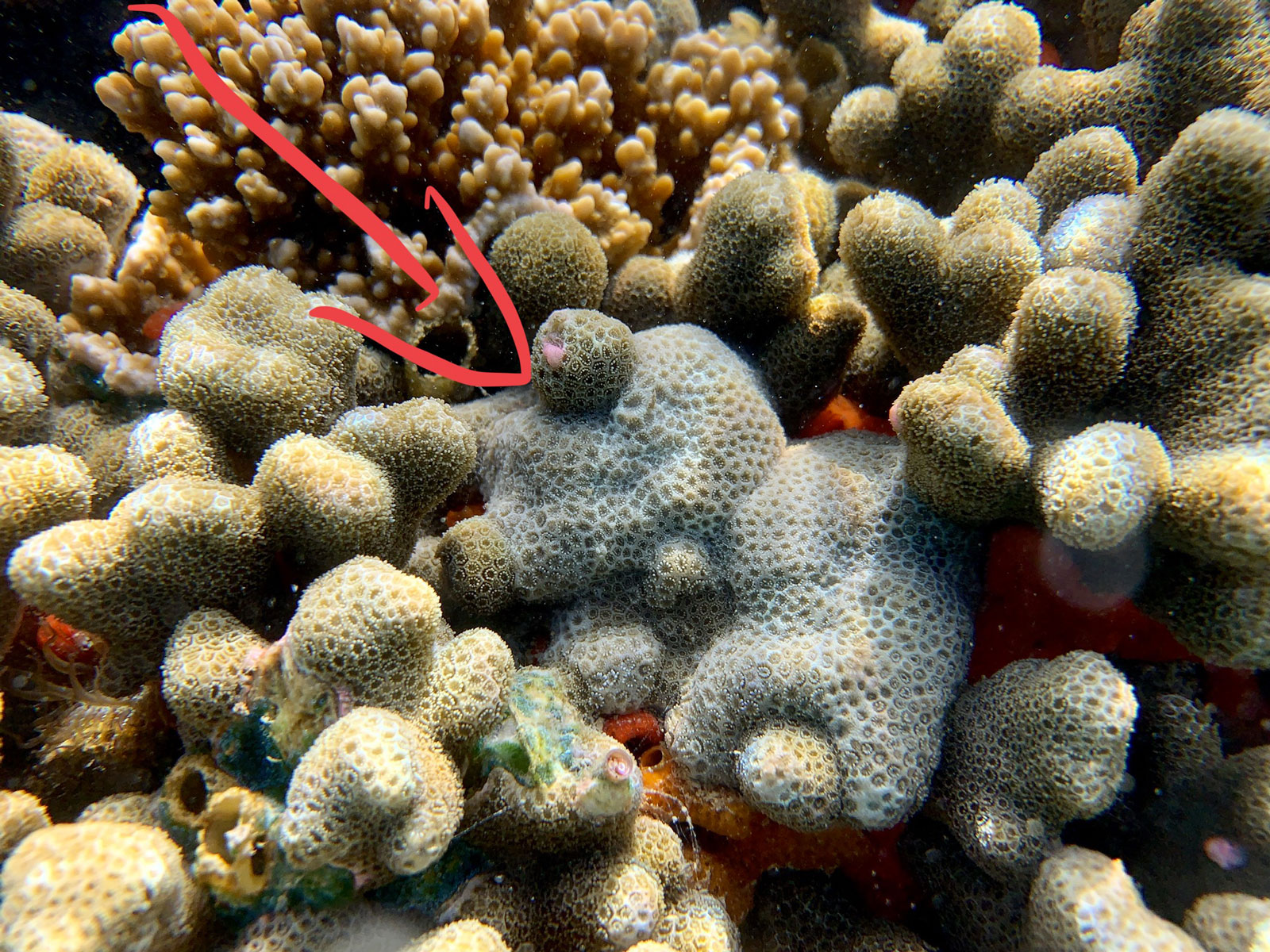
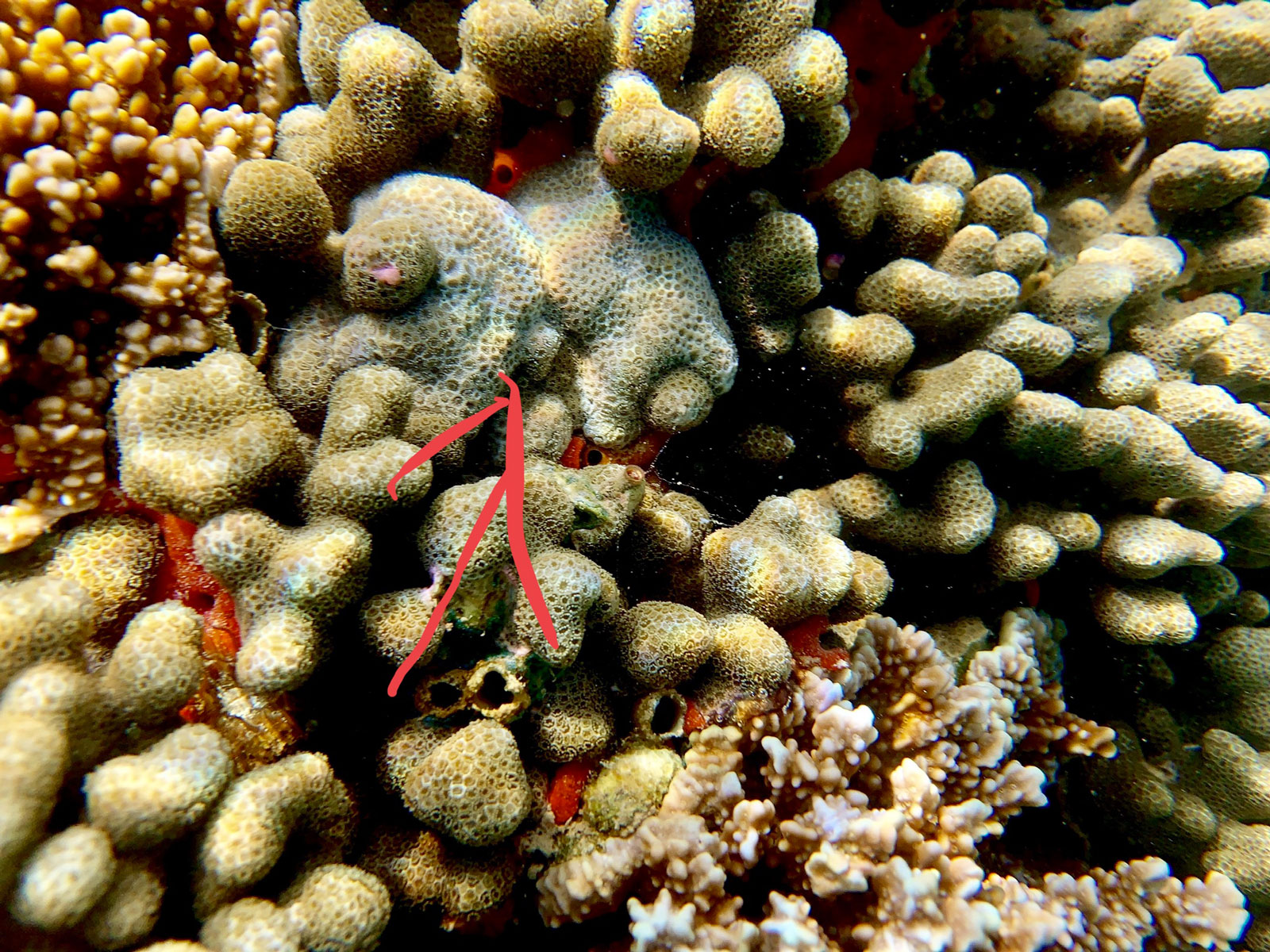
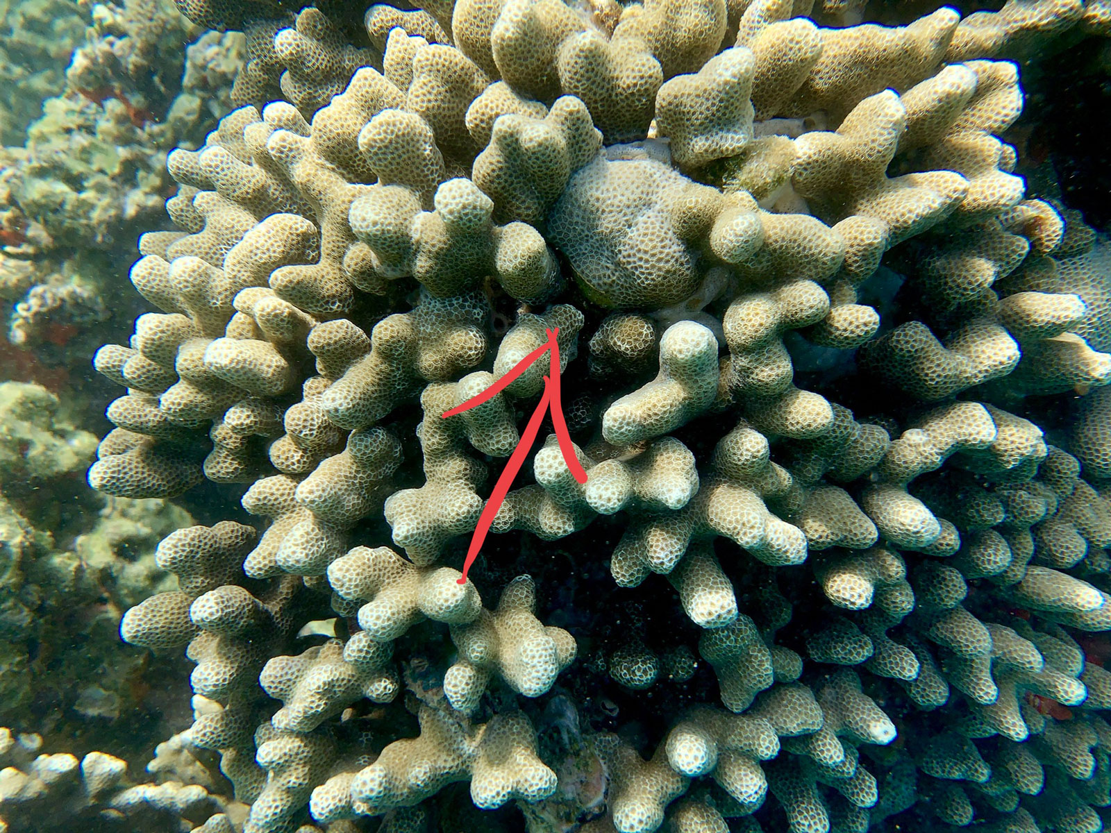
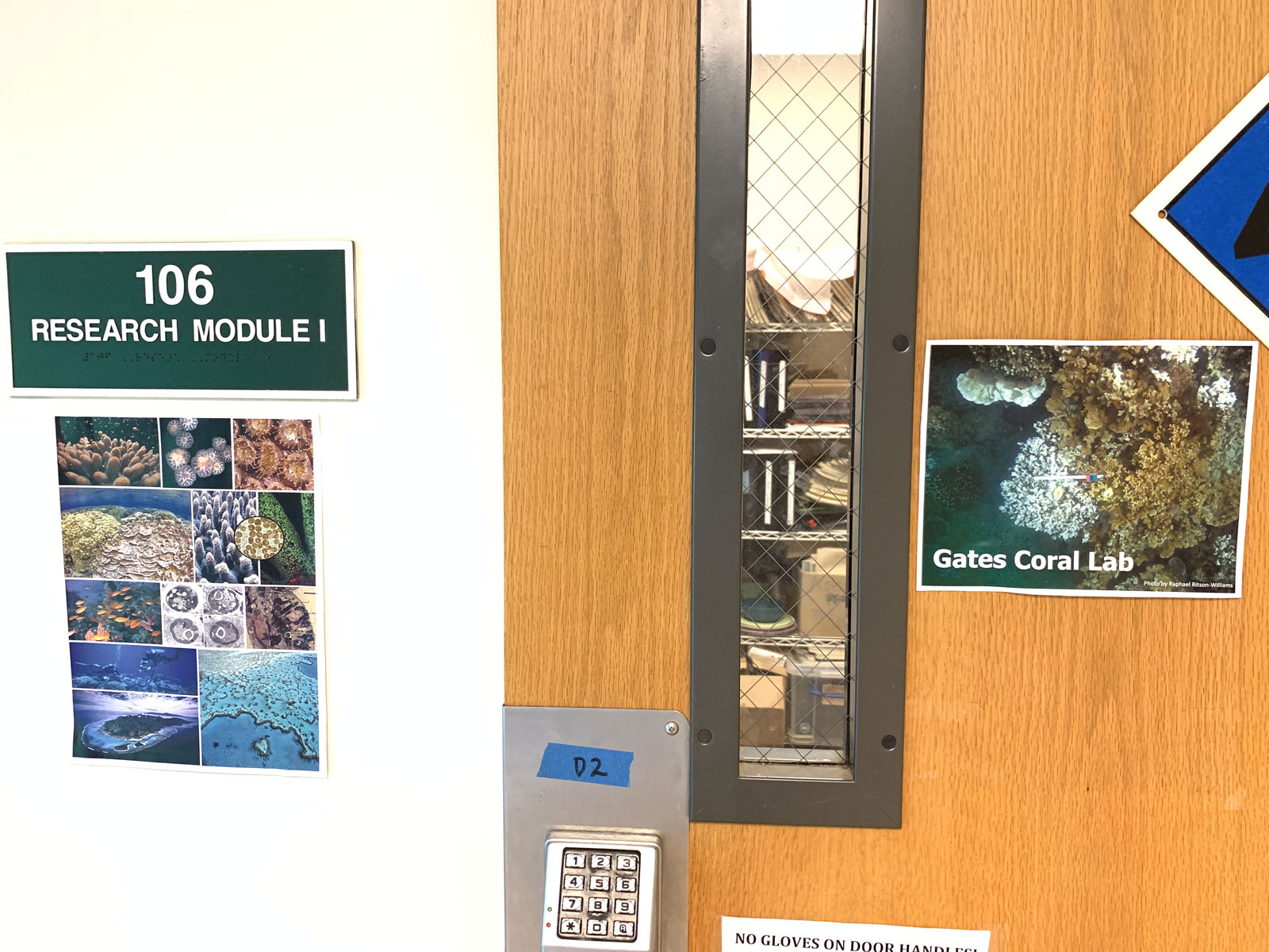
These are photos taken today from our mission in Hawaii’s Kāneʻohe Bay on Moku O Loʻe Island. Each photo displays coral showing abnormal growths which may be caused by a virus, bacteria, or water temperature changes. The scientists on site from Centre Scientifique de Monaco have taken more than 100 samples from these diseased corals and will return them to their laboratory in Monaco to continue their research.
After collecting samples of diseased corals from the sea, Drs. Dorota Czerucka and Francois Seneca must preserve the samples in various forms to ship the samples to their lab and perform their research.


We are hosted by the Hawaiʻi Institute of Marine Biology and able to use their laboratories here on Moku O Loʻe Island to preserve the samples.
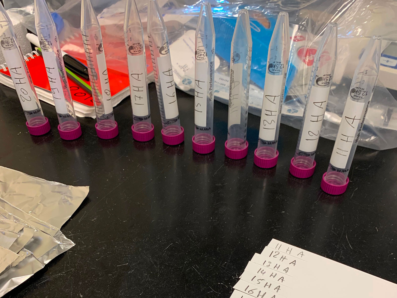
First, we use underwater paper to mark each sample, 1-10 healthy, and 1-10 with tumors.
Next, prepare the liquid nitrogen. This is one way the samples are preserved. Dropping them in the -196 degree liquid nitrogen preserves the sample exactly as it was found, so that DNA, RNA and proteins can be extracted and analyzed based on the environment in which the sample is living. The sample is cut into smaller pieces as we will preserve it multiple ways – the nitrogen is one, but we also keep some alive in sea water to transport them back to our lab in Monaco and let them continue to grow.


Next we move on to the wet lab where there is sea water pumped into tables so the samples can continue to live. First step here is to prepare these stems marked with the sample number and either H for healthy or T for tumor. The stems are placed into a tray, as shown in the photo, with the diseased coral on the left and the healthy on the right. Each sample is cut into 3 so there are more chances of a live coral being preserved, and because each will be used for a different research purpose. When we are ready to leave Hawaii, the samples are suspended on a string and floated in a bag filled with seawater and 100% oxygen, which is what they will consume to survive on their trip to Monaco.


While collecting pieces of coral, we also scrape the mucus from their surface to collect and analyze bacterial communities. Here you can see that it’s much like the coral – there is one sample of mucus taken from healthy coral and one taken from the diseased coral on the same colony. The samples are mixed with the Vortex machine which ensures the bacteria is completely mixed with the sea water, then put into centrifuge to separate the bacteria completely before preserving it in the liquid nitrogen and preparing it for shipping back to Monaco. In the photos it is easy to see the difference between the healthy mucus (clear) and the diseased mucus (brown).


To collect samples for research, the scientists must spend time in the sea searching for coral colonies that show abnormal growths. Each day, we suit up and collect our materials and head out to sea with snorkels and masks hoping to collect samples from a total of ten colonies. When one is identified, a photo is taken to document the size and GPS coordinates. Next, Francois Seneca chips away 2 pieces from the colony – one of the healthy coral and one of the diseased coral. He then stores the samples in separate bags pre labeled 1-10 for both healthy and diseased samples. Next, he repeats the process with a new colony.
Here’s a video that shows what that process looks like.
Dorota Czerucka and François Seneca from the Centre Scientifique de Monaco have collected 140 samples of both healthy and diseased corals in two locations.
The scientists are collecting samples of corals showing abnormal growths which could be tumors. The team is conducting research to understand the immune mechanism response given by the corals to this situation.


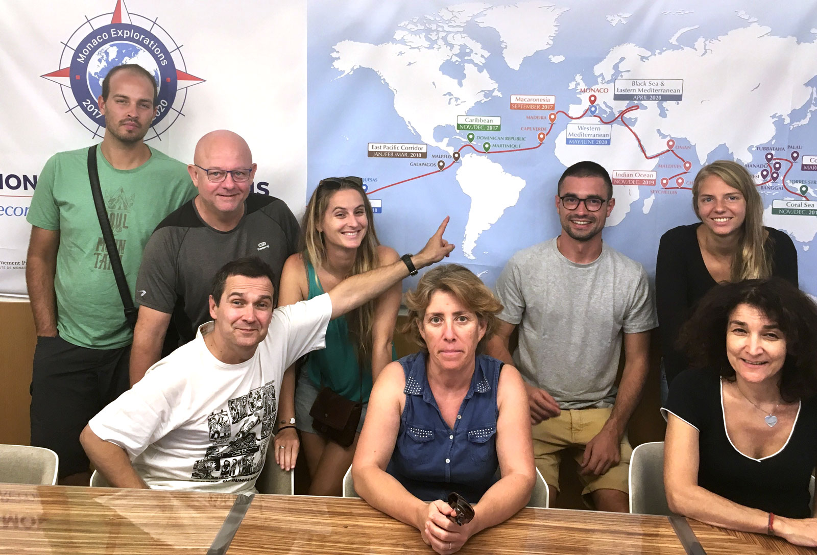
From Colombia, Switzerland and France … scientists are more than happy to be here in Malpelo a real sanctuary for marine life and biodiversity. Onboard with Monaco Explorations, their dream came true
From today, Monaco Explorations welcome a new scientific team (8 members) from MIO (Mediterranean Institute of Oceanography, Aix-Marseille University, France) to cross the Atlantic from Cape Verde to Martinique in search of Sargasso rafts. Sargasses are marine brown seaweeds. This expedition purpose is to identify the processes responsible for the proliferation of this invasive brown algae which is deleterious, depriving coastal life of light and creating big amount of organic matter in putrefaction at shore with dramatic consequences on water, air quality and health in the Caribbean.
[vc_row][vc_column][vc_column_text] [/vc_column_text][/vc_column][/vc_row]